
Ramzi Theory Explained: How It Works and Is It Accurate?
You have an early ultrasound image, a brighter area appears on one side, and now you are wondering whether it could reveal your baby’s sex.
The Ramzi Theory claims that the location of developing placental tissue during early pregnancy can predict whether a baby is a boy or a girl. According to the theory, tissue forming on the mother’s right side suggests a boy, while tissue forming on the mother’s left side suggests a girl.
However, the Ramzi Theory is not a scientifically proven method. An independent study involving 277 pregnancies found no relationship between placental location and fetal sex.
This guide explains what the Ramzi Theory claims, how people attempt to read an early ultrasound, why abdominal and transvaginal scans can cause confusion, and which medical methods offer more reliable information.
Quick answer
According to the Ramzi Theory:
- Developing placental tissue on the maternal right predicts a boy.
- Developing placental tissue on the maternal left predicts a girl.
- Central, unclear, anterior or posterior placement produces an inconclusive result unless left-right orientation is also known.
These predictions are for entertainment only. Scientific evidence does not confirm that the Ramzi Theory reliably determines fetal sex.
What Is the Ramzi Theory?
The Ramzi Theory, sometimes called the Ramzi method, is an early pregnancy sex-prediction theory based on the supposed location of developing placental tissue.
It is commonly associated with ultrasound images taken at approximately six to eight weeks of pregnancy. Followers believe the side of the uterus where placental development begins may be connected to the baby’s chromosomal sex.
The theory uses this basic rule:
| Developing tissue location | Ramzi Theory prediction |
|---|---|
| Maternal right side | Boy |
| Maternal left side | Girl |
| Centre or unclear | Inconclusive |
The method became popular because many parents want to make a prediction before fetal genital anatomy can be assessed on an ultrasound.
At six to eight weeks, however, an ultrasound is used to evaluate early pregnancy development rather than determine fetal sex. External genital appearance has not developed enough at this stage to provide a visible boy-or-girl assessment.
What does the Ramzi Theory examine?
The Ramzi Theory is supposed to examine the location of developing placental tissue, not the position of the embryo.
During early pregnancy, the placenta is still developing. People applying the theory often look for thicker or brighter tissue near one side of the gestational sac. This may be described as chorionic or trophoblastic tissue.
The problem is that a non-professional may confuse this tissue with:
- The embryo
- The yolk sac
- The uterine wall
- An ultrasound shadow
- A bright tissue boundary
- An image artefact
An early ultrasound is a medical image. Only a qualified sonographer, radiologist or maternity care professional should interpret its clinical findings.
Is it based on the baby’s position?
No. The embryo’s position is not the feature that the theory claims to use.
The embryo may appear on one side of the gestational sac while the developing placental tissue appears somewhere else. Looking at where the embryo seems to be sitting can therefore produce a completely different guess.
Common mistake:
The small structure inside the gestational sac is usually the embryo or yolk sac—not necessarily the developing placenta.
Ramzi Theory Results at a Glance
The following table shows what different locations mean under the theory and when a responsible result should be considered inconclusive.
| Observed location | Traditional Ramzi result | Important limitation |
|---|---|---|
| Maternal right | Boy | The theory is not scientifically validated |
| Maternal left | Girl | The theory is not scientifically validated |
| Centre or midline | Inconclusive | There is no clear left-right placement |
| Anterior | Inconclusive without left-right information | Anterior means the front of the uterus |
| Posterior | Inconclusive without left-right information | Posterior means the back of the uterus |
| Fundal | Inconclusive without left-right information | Fundal means the top of the uterus |
| Unclear tissue | Inconclusive | The visible area may not be placental tissue |
| Screen-left or screen-right only | Inconclusive | Screen position may not match maternal anatomy |
| Cropped or rotated image | Inconclusive | Orientation labels may be missing |
| Twin pregnancy | Inconclusive | A single left-right rule cannot assess multiple sacs or placentas reliably |
Anterior, posterior, and fundal are not alternative words for left and right. An anterior placenta is attached toward the front wall of the uterus, a posterior placenta is toward the back, and a fundal placenta is near the top.
How People Apply the Ramzi Theory
You can use the following process for a light-hearted prediction, but it should not replace professional medical information.
1. Check the gestational week
The Ramzi Theory is most commonly discussed in relation to scans taken at around six to eight weeks.
The exact gestational age matters because early pregnancy structures change quickly. However, having a scan during the commonly discussed period does not make the theory scientifically reliable.
2. Confirm the scan type
Check whether the ultrasound was:
- Transvaginal
- Transabdominal
- A combination of both
- Unknown
This information may appear on the report, or the clinic may be able to confirm it.
3. Find the developing placental tissue
Followers of the theory look for an area of thicker or brighter tissue attached near the gestational sac.
Do not simply follow the embryo’s position. The embryo and developing placenta are separate structures.
4. Confirm anatomical orientation
This is the most important—and most frequently overlooked—step.
You need to know whether the tissue is on the mother’s anatomical right or left. Knowing only that it appears on the left or right side of a saved image is not enough.
5. Choose right, left or inconclusive
Select maternal right or maternal left only when anatomical orientation has been confirmed.
Use inconclusive when:
- The image orientation is unknown.
- The tissue appears central.
- The image is unclear or cropped.
- You only know the position on the screen.
- The placenta is described only as anterior, posterior, or fundal.
- Different people identify different areas.
- The scan contains multiple gestational sacs.
6. Read the entertainment-only prediction
According to the theory:
- Maternal right suggests a boy.
- Maternal left suggests a girl.
The result remains a guess, even when the image orientation appears clear.
Abdominal vs Transvaginal Ultrasound
Early pregnancy ultrasound may be performed across the abdomen or with a transvaginal probe.
| Feature | Abdominal ultrasound | Transvaginal ultrasound |
|---|---|---|
| How it is performed | A probe moves across the lower abdomen | A covered probe is inserted into the vagina |
| Early pregnancy image | May provide a wider pelvic view | Often provides clearer early detail |
| Does scan type confirm left and right? | No | No |
| Must the image always be flipped? | No | No |
| Best orientation source | Sonographer, report or image markers | Sonographer, report or image markers |
| Ramzi Theory reliability | Unproven | Unproven |
Does an abdominal ultrasound need to be flipped?
Not automatically.
Many online guides claim that every abdominal ultrasound must be flipped before applying the Ramzi Theory. This rule is too simplistic.
The appearance of the image may depend on:
- The scanning plane
- The direction of the probe
- The probe marker
- The ultrasound machine’s display convention
- The position of the uterus
- Whether the image was rotated or cropped
- Whether the original labels remain visible
You should not reverse a prediction solely because the scan was abdominal.
Are transvaginal ultrasound images mirrored?
You should not assume that every transvaginal image follows one universal mirrored or non-mirrored format.
A sonographer can capture sagittal, transverse, and other views by changing the probe’s direction and angle. Clinical orientation also depends on the probe marker and scanning plane.
The safest question to ask your clinic is:
“Can you show me which side of this image corresponds to my anatomical right and left?”
You are not asking the sonographer to support the Ramzi Theory. You are simply asking for help understanding the image orientation.
Why Ultrasound Orientation Causes Confusion
The biggest practical weakness of the Ramzi Theory is not simply identifying a bright area. It is proving which side of the mother’s body that area represents.
Screen-left does not always mean maternal-left
A saved ultrasound image represents a specific slice through the body.
Without visible labels or information about the scanning plane, screen-left could correspond to a different anatomical direction. It should not automatically be interpreted as the mother’s left side.
This explains why two people can examine the same image and provide opposite predictions. One may interpret screen-left as maternal-left, while the other assumes the image needs to be reversed.
Cropped images can remove essential information
An ultrasound image posted online may have been:
- Cropped around the gestational sac
- Rotated to make it easier to view
- Flipped by a phone or editing application
- Photographed from a printed scan
- Saved without its orientation labels
- Captured from a moving ultrasound video
The brighter tissue may appear obvious while the anatomical direction remains completely unknown.
Inconclusive can be the most accurate answer
A responsible Ramzi predictor should not force every scan into a boy or girl result.
When orientation or tissue location cannot be confirmed, inconclusive is the most honest answer.
Can You Use the Ramzi Theory at 5, 6, 7, 8 or 9 Weeks?
People attempt Ramzi predictions at different stages of early pregnancy, but image clarity can vary considerably.
| Pregnancy week | What may be visible | Main Ramzi limitation |
|---|---|---|
| 5 weeks | A small gestational sac and possibly a yolk sac | Developing tissue may be too early or unclear |
| 6 weeks | Gestational sac, yolk sac and a small embryo may be visible | Tissue and orientation can still be difficult to identify |
| 7 weeks | Early structures are usually more noticeable | A clearer image does not validate the theory |
| 8 weeks | The embryo and surrounding tissue may be easier to see | Bright tissue can still be misidentified |
| 9 weeks | Early pregnancy development is generally more distinct | Placental side remains an unproven predictor |
| 10 weeks or later | More fetal development may be visible | The Ramzi Theory remains scientifically unsupported |
Gestational dating, ultrasound equipment, scanning technique, body position, and whether the scan is abdominal or transvaginal can all affect what is visible.
Do not use an online Ramzi interpretation to assess pregnancy viability, growth, implantation or the cause of symptoms.
Is the Ramzi Theory Accurate?
No reliable evidence shows that the Ramzi Theory can accurately predict fetal sex.
The most relevant independent study evaluated 277 pregnancies. The placenta was positioned on the right in 89 cases, and only 51% of those fetuses were male. It was positioned on the left in 110 cases, and 57% of those fetuses were female. The researchers concluded that there was no relationship between placental location and fetal sex.
What about the widely repeated 97% accuracy claim?
Some websites repeat claims that the theory is more than 90% or even 97% accurate.
These figures should not be presented as established medical facts. The original material associated with the theory was not published as a conventional, transparent peer-reviewed clinical study that could be independently evaluated and reproduced.
A high percentage appearing on many websites does not become reliable simply because it is repeated.
Why does peer review matter?
Peer review helps independent specialists examine:
- How the study participants were selected
- How placental location was defined
- How scan orientation was confirmed
- Whether unclear images were excluded
- Whether assessors already knew the fetal sex
- Whether the statistical analysis was appropriate
- Whether another research team could reproduce the findings
Without clear methods and independent replication, an accuracy claim may be affected by bias, selective reporting, or inconsistent interpretation.
Why do many parents say it worked?
A person may genuinely receive a correct Ramzi prediction. That does not prove the method caused or predicted the correct result.
A basic boy-or-girl guess has only two common outcomes. Many predictions will therefore match the eventual result by chance.
People may also remember successful predictions more clearly than unsuccessful ones or share correct results more often in online groups.
Evidence rating
Scientific support: Insufficient
Independent validation: Not established
Clinical use: Not recommended
Best use: Entertainment only
Medical confirmation required: Yes
Common Reasons Ramzi Predictions Go Wrong
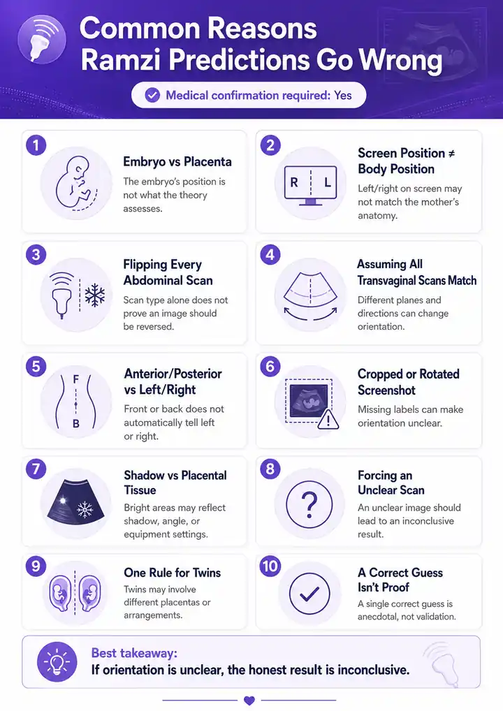
Looking at the embryo instead of placental tissue
The embryo’s position is not the feature the theory claims to assess.
Assuming screen position equals body position
Screen-right and screen-left may not represent the mother’s anatomical right and left.
Automatically flipping every abdominal scan
Scan type alone does not tell you whether the saved image should be reversed.
Assuming every transvaginal scan has the same orientation
Transvaginal images can be captured in different planes and directions.
Confusing anterior or posterior with left or right
Anterior means front, while posterior means back. Neither term provides a left-right result by itself.
Using a cropped or rotated screenshot
The image may no longer contain the labels needed to understand its orientation.
Mistaking a shadow for placental tissue
Ultrasound brightness can be affected by tissue boundaries, probe angle, depth, and equipment settings.
Forcing an unclear scan into a result
An unclear image should produce an inconclusive result—not a confident boy or girl prediction.
Applying one prediction to twins
Multiple pregnancies may involve different sacs, placentas, and chorionic arrangements. A single left-right rule cannot assess them responsibly.
Treating a correct guess as proof
One correct prediction is an anecdote, not scientific validation.
A Practical Ramzi Theory Example
Imagine that you have a seven-week transvaginal ultrasound.
A brighter area appears on the right side of the saved image. At first, it may look like a clear boy result.
Before making that prediction, you would need to know:
- Does image-right represent maternal right?
- Was the scan captured in a transverse or sagittal plane?
- Has the image been rotated, cropped, or flipped?
- Is the brighter area actually developing placental tissue?
Suppose the image has no visible orientation labels and the clinic has not confirmed which side represents maternal right.
The appropriate result would be:
Inconclusive: the image shows screen position, but maternal orientation has not been confirmed.
This answer may be less exciting than a forced prediction, but it is more honest and useful.
Ramzi Theory vs Nub Theory vs Skull Theory
The Ramzi Theory is only one of several popular prenatal prediction methods.
| Method | What it examines | Common timing | Scientific support |
|---|---|---|---|
| Ramzi Theory | Supposed location of developing placental tissue | Around 6–8 weeks | Insufficient |
| Nub Theory | Angle of the developing genital tubercle | Around 11–14 weeks | Limited; casual image guesses remain uncertain |
| Skull Theory | Shape of the fetal skull | Usually later in the first or second trimester | No dependable support for individual prediction |
| Medical ultrasound | Visible external genital anatomy | Commonly during the 18–22-week anatomy scan | Clinically established when anatomy is visible |
| Genetic testing | Chromosomal information | Depends on the test | Clinically established within each test’s purpose and limitations |
The standard pregnancy ultrasound is usually performed between 18 and 22 weeks. Its main purpose is to examine fetal anatomy, development and pregnancy health—not simply to identify fetal sex.
Reliable Ways to Learn a Baby’s Sex
Several medically established methods may provide information about fetal sex. However, this is not always their primary purpose.
| Method | Typical timing | Main purpose | Important limitation |
|---|---|---|---|
| Cell-free DNA screening or NIPT | From around 10 weeks | Screening for certain chromosome conditions | It is a screening test, not a diagnosis |
| Anatomy ultrasound | Usually 18–22 weeks | Evaluating fetal anatomy and development | Visibility depends on fetal position and image quality |
| Chorionic villus sampling | First trimester | Diagnostic genetic or chromosome testing | Invasive and performed for clinical reasons |
| Amniocentesis | Usually during the second trimester | Diagnostic genetic or chromosome testing | Invasive and performed for clinical reasons |
| Preimplantation genetic testing | Before embryo transfer during IVF | Testing embryos for certain chromosomal or genetic findings | Only available as part of IVF treatment |
Cell-free DNA screening or NIPT
Cell-free DNA screening can be performed from approximately 10 weeks of pregnancy. It screens for certain chromosomal conditions and sex-chromosome differences. It remains a screening test, so a positive result may require diagnostic confirmation.
Anatomy ultrasound
A standard anatomy ultrasound is generally performed between 18 and 22 weeks. The sonographer may be able to identify external genital anatomy when the baby’s position and image quality allow it.
CVS and amniocentesis
Chorionic villus sampling and amniocentesis are diagnostic tests that examine cells obtained from placental tissue or amniotic fluid. These procedures are used to investigate genetic or chromosomal conditions and should not be undertaken merely for a casual sex prediction.
Preimplantation genetic testing
Preimplantation genetic testing is used during IVF to examine embryos for specified genetic or chromosomal findings before embryo transfer. Some forms include analysis of the X and Y chromosomes.
Speak with your maternity care provider or a genetic counsellor to understand which screening or diagnostic options are appropriate for your pregnancy.
Should You Trust a Ramzi Theory Result?
You can enjoy a Ramzi prediction as a light-hearted pregnancy activity, but you should not treat it as confirmation.
Do not base the following on a Ramzi result:
- Medical or prenatal-testing decisions
- Major emotional expectations
- Decisions influenced by a preference for one sex
- Pregnancy announcements presented as medically confirmed
- Expensive or non-returnable purchases
- Interpretation of pain, bleeding or unusual ultrasound findings
Your ultrasound contains important medical information that goes far beyond a gender-prediction game. Ask your healthcare professional when you have questions about pregnancy location, viability, fetal development, or placental position.
Try Another Traditional Gender Predictor
If you enjoy comparing traditional prediction methods, you can also try our Mayan Calendar Gender Calculator for another just-for-fun result.
The Mayan calendar method is also a traditional entertainment activity. It cannot medically determine fetal sex and should not influence pregnancy-related decisions.
Final Takeaway
The Ramzi Theory offers an early and entertaining pregnancy guess, but it cannot medically confirm fetal sex.
The traditional rule is simple: maternal right supposedly predicts a boy, while maternal left supposedly predicts a girl. The difficult part is confirming which anatomical side an ultrasound image shows and correctly identifying the developing placental tissue.
Even when those details are known, scientific research has not established placental location as a reliable predictor of fetal sex.
Use the theory for fun, accept an inconclusive result when information is missing, and rely on qualified prenatal care for dependable answers.
FAQs
What is the Ramzi Theory in simple terms?
The Ramzi Theory claims that developing placental tissue on the mother’s right predicts a boy, while tissue on the mother’s left predicts a girl. Scientific evidence does not confirm that it works reliably.
Which side means boy in the Ramzi Theory?
According to the theory, developing placental tissue on the maternal right suggests a boy. Screen-right should not be treated as maternal right unless anatomical orientation is confirmed.
Which side means girl in the Ramzi Theory?
According to the theory, developing placental tissue on the maternal left suggests a girl. This remains an entertainment-only prediction.
Is the Ramzi Theory scientifically proven?
No. An independent study involving 277 pregnancies found no relationship between placental location and fetal sex.
How accurate is the Ramzi Theory?
Its dependable accuracy has not been established. Widely repeated accuracy percentages should not be treated as proven clinical statistics.
Can the Ramzi Theory work at five weeks?
There is no evidence that it works reliably at five weeks. This is also a very early stage, so the structures people attempt to identify may be small or unclear.
Is six weeks too early for a Ramzi prediction?
Six weeks is commonly discussed by followers of the theory, but that timing does not make the prediction medically reliable.
Medical Disclaimer
This article is provided for general education and entertainment only. It does not offer medical advice, interpret ultrasound findings, or confirm fetal sex. Do not use a Ramzi Theory prediction to make medical, emotional, financial or pregnancy-related decisions.
Speak with a qualified midwife, sonographer, obstetrician, radiologist, or other maternity care professional about your individual pregnancy.
Seek prompt medical attention if you experience significant bleeding, severe abdominal pain, fainting or other concerning pregnancy symptoms.